The human body is full of subtle indicators that can betray underlying medical illness, and your pupils are no exception. Pupil size and response can be an early warning sign of neurological disease, cognitive impairment, and even head injury. Medical technology has advanced to the point where pupil testing has reached the most accurate levels ever, due to the use of neurological devices like the NPi (Neurological Pupil Index). But what do these tiny changes in your pupil actually reveal about brain health? Let’s explore the fascinating connection between pupil behavior and neurological function. Pupil behavior is an important marker of neurological function that can give rise to earlier detection and treatment, ultimately improving patient outcomes and quality of life.
The Science of Pupil Reactivity
Pupillary response is regulated by the autonomic nervous system and governed by the brainstem. It comprises two main reflexes: the light reflex and the sound reflex.
- Constriction (Miosis): The pupil of the eye is narrowed by the presence of light or when viewing objects at a near distance. The parasympathetic nervous system carries out this reflex and is achieved through the sphincter pupillae muscle.
- Dilation (Mydriasis): The pupil enlarges in dim light or as a response to the brain’s stimulus for an arousal response, e.g., excitement or stress. This is under sympathetic nervous system control and is regulated by the dilator pupillae muscle.
Any change in these reactions can suggest possible neurological disorders, and pupil tests thus constitute a significant part of neurological examination. These reactions are essential to visual adaptation and can yield valuable information about the integrity of the neural pathways for autonomic function.
Pupil Exam: A Window into Neurological Health
Pupil testing is a rapid, safe, and simple way of assessing brain function. Neurologists, ophthalmologists, and emergency medical staff use pupil testing to detect:
- Brain Trauma & Concussions – Asymmetrical or delayed pupil reaction could be a sign of head trauma. The changes can be indicative of damage to the nerve tracts that control pupil size and are useful for determining the severity of injury.
- Stroke & Brain Hemorrhages – Sudden changes in pupil size may suggest serious cerebrovascular diseases. Early testing of pupil response may allow for early detection and treatment of stroke and enhance patient outcomes.
- Neurodegenerative Disorders – Neurodegenerative diseases like Alzheimer’s and Parkinson’s exhibit distinctive pupil reactivity patterns. Mild changes in pupillary response are the first indications of these long-term diseases, and intervention and treatment can be initiated at a young age.
- Intracranial Pressure (ICP) Changes – Pupillary dilation can be an indication of increasing brain pressure that can sometimes have a need for urgent treatment. Monitoring of pupil size and reactivity can be helpful in intensive care units for the detection and management of possibly life-threatening increases in ICP.
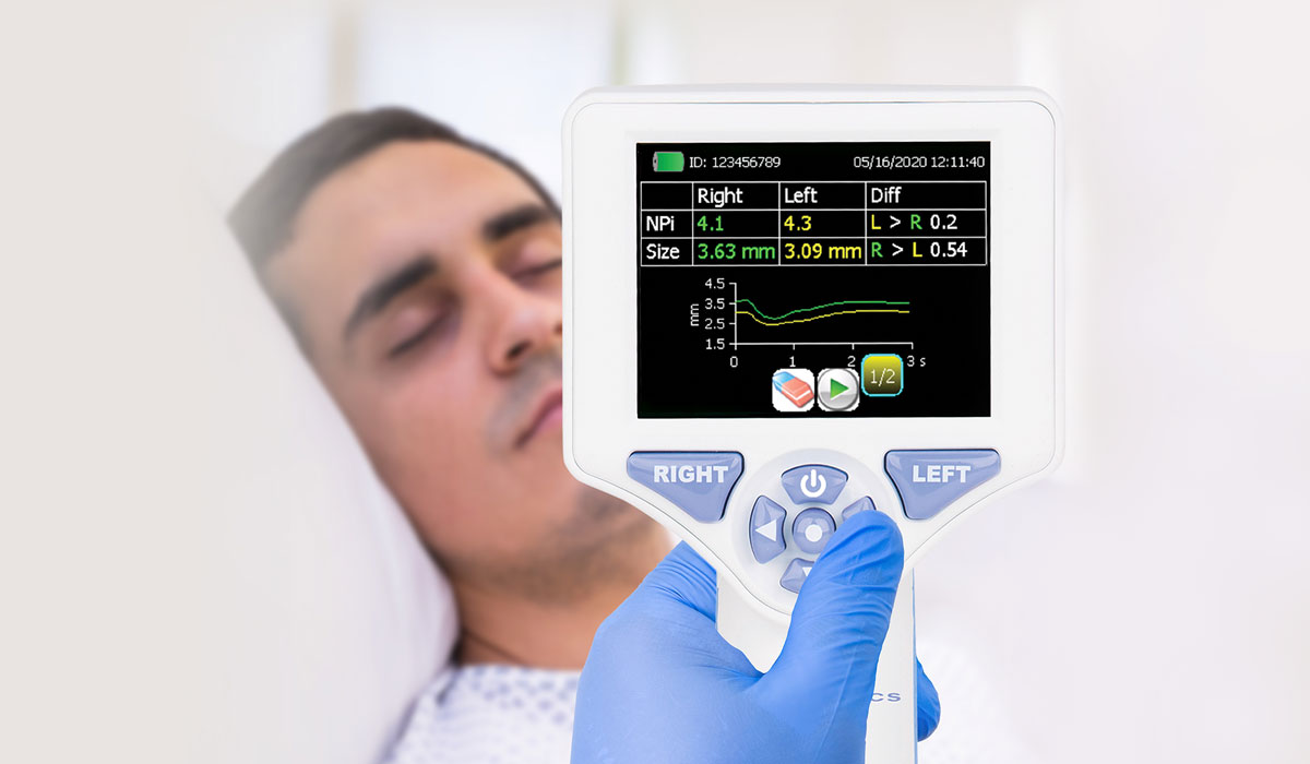
How NPi Improves Pupil Assessment
The Neurological Pupil Index (NPi) is a novel method of pupil response assessment. Traditional assessment relies on subjective observation, whereas the NPi uses objective measurement to provide standardized results. The NPi pupillometer measures pupil diameter in real time, allowing physicians to:
- Catch early neurological changes: In providing precise, quantitative data, the NPi is able to identify subtle changes in the pupil response that a visual inspection might miss.
- Monitor the development of brain injury: Regular and impartial tests provide accurate measurement of change, allowing treatment benefit and patient outcome to be measured.
- Enhance neuro exam reliability: The NPi reduces variability with subjective tests, providing more reproducible and consistent findings, essential for accurate evaluation and treatment planning.
What Different Changes in Pupils Might Mean
Other pupil abnormalities may point to other underlying neurological conditions. Some of the most common pupil changes and their potential implications include:
| Pupil Change | Possible Cause |
| Asymmetrical pupils (Anisocoria) | Brain injury, stroke, nerve injury, Horner’s syndrome, third cranial nerve palsy |
| Sluggish reaction to light | Concussion, drug action, neurodegenerative illness, optic neuritis, elevated intracranial pressure |
| Fixed and dilated pupils | Severe head trauma, cardiac arrest, severe hypoxia, certain drugs or toxins |
| Excessive narrowing (Miosis) | Opioid use, brainstem trauma, pontine hemorrhage, some drugs |
| Abrupt pattern changes (Hippus) | Normal fluctuation or early neurologic impairment, certain neurological syndromes, early phase of brain injury |
Recent Studies of Pupillary Response and Cognitive Well-being
Recent research has proven a close association between cognitive capacity and pupil reactivity. Researchers are currently employing pupil response as a biomarker for the detection of early cognitive decline and dementia. Some of the findings are:
- Alzheimer’s Detection: Slowed pupillary constriction and re-dilation are manifestations in early patients with Alzheimer’s. This means that abnormalities of the pupillary light reflex are potentially an indicator of early neurodegenerative change in Alzheimer’s disease.
- Mental Effort and Attention: Mental effort is directly correlated with pupil size—larger pupil sizes are associated with larger mental effort. It is therefore a non-invasive quantification of cognitive effort and fatigue and offers insight into brain function under various tasks.
- Autonomic Nervous System Disorders: Autonomic nervous system disorders of the multiple system atrophy type impair normal pupil dynamics. Pupil reactivity changes are helpful for differential evaluation and follow-up of autonomic nervous system disorders, which typically manifest as pupillary dysfunction.
The Future of Pupillary Examination in Medicine
With advancements in technology, pupil tests are now becoming a standard part of regular medical checkups. Technological innovations in neurological equipment such as the NPi pupillometer are improving the accuracy of examination, resulting in quicker intervention and better patient outcomes. Some of the future applications that are likely to emerge include:
- AI-Based Pupil Analysis – Pupil data is analyzed in real time by machine learning software for identifying diseases during their early development stages. Such applications may detect faint patterns and anomalies that are hard to identify by humans, allowing for earlier detection of neurological diseases.
- Wearable Pupil Monitoring Devices – Neurological health can be tracked using smart glasses or headsets. This technology can provide real-time monitoring of pupil responses in a non-invasive way, allowing for early detection of change and proactive maintenance of neurological health.
- Pupil Biometrics to Enable Precision Medicine – Pupil response measures have the potential to support personalized therapy in neurodegenerative diseases. With heterogeneity in pupil reactivity data in patients, clinicians will be able to formulate better-informed and enhanced treatment plans, maximizing patient outcomes.
Conclusion
Your pupils are not merely windows to the soul, but potent signals of brain function. From concussions to dementia, pupil examination gives us profound neurological information. Improved measurement of pupil diameter and technology such as the NPi pupillometer are transforming medical examinations. With further research, pupillary response could become the most critical metric for the evaluation and preservation of brain health. Not only will this technology refine neurological evaluation with greater accuracy and efficiency, but it will open the door to preventative interventions and customized care and ultimately provide improved quality of life for neurologically compromised individuals.